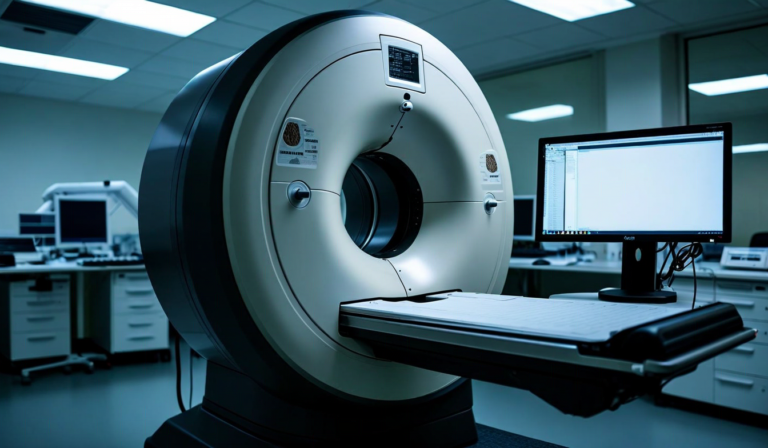
Single-photon emission computed tomography occupies a complicated position in the nuclear medicine landscape. It images at lower spatial resolution than PET, uses gamma cameras that have changed relatively little in fundamental design since the 1980s, and lacks the absolute quantification that time-of-flight PET enables. Yet SPECT handles a broader set of clinical indications than PET - myocardial perfusion imaging, DaTscan for Parkinson's diagnosis, bone scanning, renal function assessment, and hepatobiliary studies among them. For most hospitals, SPECT volume exceeds PET volume by a factor of three to four. Deep learning is changing what that large installed base can diagnostically deliver.
The Physical Limitations Deep Learning Targets
SPECT image quality is degraded by several well-characterized physical phenomena. Attenuation - the absorption of gamma photons by patient tissue before they reach the detector - reduces apparent tracer concentration in deep structures and creates artifactual defects, particularly in myocardial perfusion imaging where diaphragm and breast tissue commonly produce false-positive inferior and anterior wall defects respectively.
Scatter - gamma photons that change direction after Compton interaction with tissue before reaching the detector - adds a diffuse background that reduces image contrast. Partial volume effects, as with PET, cause small structures to appear larger and with lower apparent activity. Collimator-detector distance causes spatial resolution to degrade with increasing depth, unlike PET where resolution is largely uniform across the field of view.
Conventional SPECT reconstruction algorithms address attenuation using CT-based attenuation correction maps, and scatter through energy window-based subtraction methods. Neither fully compensates for these effects, and both introduce their own artifacts when the CT and SPECT acquisitions are misregistered or when the patient moves between them. Deep learning approaches these corrections differently - by learning the relationship between degraded and corrected images directly from large training datasets, rather than solving the physical correction problem analytically.
Deep Learning Reconstruction: What Changes
The most direct application of deep learning to SPECT is post-reconstruction image enhancement. A convolutional neural network trained on paired low-count and high-count SPECT acquisitions learns to predict the high-quality image from the degraded one. The clinical implication is significant: if a model can reliably reconstruct a high-quality image from a half-dose or quarter-dose acquisition, dose reduction or scan time reduction becomes feasible without sacrificing diagnostic quality.
Published validation studies in myocardial perfusion SPECT show that deep learning-enhanced images from 50% count-rate acquisitions match the diagnostic quality of standard full-count studies as assessed by expert readers, with defect detectability maintained. For bone scan applications, where the primary diagnostic task is identifying focal areas of increased osteoblastic activity, similar results have been reported with count reduction factors of 40-50%.
A separate branch of deep learning reconstruction targets quantitative accuracy rather than visual quality. Iterative reconstruction algorithms (OSEM - ordered subsets expectation maximization) remain the clinical standard, but the number of iteration/subset combinations used clinically is often chosen for visual appearance rather than quantitative accuracy. Deep learning reconstruction methods trained on high-iteration reference reconstructions can achieve comparable quantitative accuracy with fewer iterations, reducing computation time and creating more reproducible quantitative outputs - relevant for dosimetry calculations and treatment response monitoring.
The Myocardial Perfusion Case Study
Myocardial perfusion imaging represents the highest-volume SPECT application and the one with the most published AI literature. The diagnostic task - identifying relative reduction in perfusion in ischemic myocardium - is well-defined, and the output classification (normal vs. abnormal by territory and severity) is standardized through ASNC and ACC/AHA guidelines. This combination makes it a favorable target for deep learning classification models.
Neural network-based MPI interpretation models trained on large institutional datasets (typically 5,000-20,000 studies) achieve sensitivity and specificity for obstructive CAD (defined against invasive coronary angiography or FFR) comparable to experienced nuclear medicine physicians. Importantly, these models handle the most common artifact sources - soft tissue attenuation artifacts - more consistently than human readers, whose threshold for calling an artifact varies with experience and individual reading style.
The limitation is what the model cannot see: clinical context. A patient with known prior myocardial infarction, a history of CABG with known graft occlusion, or a presentation that clinically favors non-obstructive disease requires information the model does not have access to. AI-assisted MPI reads should always be presented alongside clinical data in the final report, a workflow step that remains entirely under physician control.
DaTscan and Parkinson's Differentiation
SPECT with Ioflupane I-123 (DaTscan) for dopamine transporter imaging represents a more specialized but diagnostically important application. The clinical task is distinguishing idiopathic Parkinson's disease and related parkinsonian syndromes from essential tremor and drug-induced parkinsonism - conditions that can be clinically indistinguishable in early stages but have very different prognoses and treatment implications.
Quantitative DaTscan analysis uses the striatal binding ratio (SBR), comparing caudate and putamen uptake to a cerebellar reference region. Visual assessment by experienced readers remains the reference standard, but intra- and inter-reader variability in borderline cases is substantial. Deep learning classification of DaTscan studies - trained on studies with confirmed clinical diagnoses - shows sensitivity of 93-95% for Parkinsonism in published series, with specificity of 88-92%. For borderline cases, agreement between AI output and initial physician interpretation ranges from 75-85%, with the AI output sometimes reflecting the consensus second-reader diagnosis rather than the initial read.
DOTATATE and Neuroendocrine Tumor Staging
Ga-68 DOTATATE PET/CT has largely replaced In-111 octreotide SPECT for neuroendocrine tumor (NET) staging in centers that have access to a cyclotron. But many community hospitals and smaller cancer centers still rely on In-111 octreotide SPECT or Tc-99m HYNIC-TOC SPECT for NET staging and treatment response monitoring. In these settings, deep learning enhancement of SPECT image quality has direct clinical impact - improved lesion contrast and reduced noise allows detection of smaller liver and lymph node metastases that influence staging and treatment decisions.
As we describe in our article on thyroid cancer staging with SPECT, the relationship between SPECT image quality and therapeutic decision-making is most acute in scenarios where the detection of a small lesion changes the treatment plan - a common situation in neuroendocrine oncology.
The Argument for SPECT Investment
The practical case for investing in deep learning-enhanced SPECT is stronger than the academic literature alone suggests. PET infrastructure requires cyclotron access or reliable radiopharmacy delivery, specialized installation costs, and scanner acquisition costs that are 2-3x those of modern SPECT/CT systems. Deep learning tools that meaningfully improve SPECT diagnostic performance represent a higher-value investment per dollar for community hospitals than PET adoption - particularly for the cardiac, neurological, and general oncology applications that dominate SPECT volume. The data supporting this argument is accumulating, even if it receives less attention than the PET AI literature.
NucliVision Supports SPECT and PET
Our platform applies deep learning enhancement across both SPECT and PET modalities, working within your existing gamma camera and PET/CT infrastructure.
View Solutions Request a Demo